Edmund Optics®

Knowledge Center

 Verified library of trusted technical resources created by our 240+ global engineers.

Filter
Search Results for: New Microscopy (162)

Multiphoton Microscopy

Multiphoton microscopy is ideal for capturing high-resolution 3D images with reduced photobleaching and phototoxicity compared to confocal microscopy.

View Now Add to saved content

Confocal Microscopy

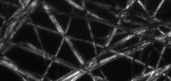
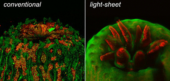
Confocal microscopy provides high resolution, elimination of out-of-focus glare due to spatial filtering, and reduction of light-induced damage to the sample.

View Now Add to saved content

Open-Source DIY Microscopy

Accessible, Flexible Technology Supported by a Community of Users

View Now Add to saved content

Light Sheet Microscopy

Light sheet fluorescence microscopy uses a 2D laser sheet to illuminate a thin slice of the sample and excite fluorescence, reducing phototoxicity and damage.

View Now Add to saved content

Discovering DIC Microscopy

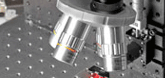
Join Stefaan Vandendriessche, Sales and Applications Engineer, as he demonstrates a Differential Interference Contrast (DIC) microscope built from stock components found in the Edmund Optics catalog.

View Now Add to saved content

Fluorescence Microscopy

View Now Add to saved content

Confocal Microscopy

View Now Add to saved content

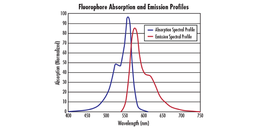
Fluorophores and Optical Filters for Fluorescence Microscopy

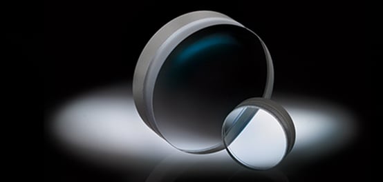
Want to know more about fluorophores and optical filters for fluorescence microscopy? Find out more information and in stock optical filters at Edmund Optics.

View Now Add to saved content

The Importance of Numerical Aperture for Imaging and Microscopy

LAB SERIES: Join us for a discussion about the importance of numerical aperture for imaging and microscopy applications.

View Now Add to saved content

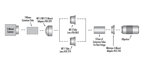
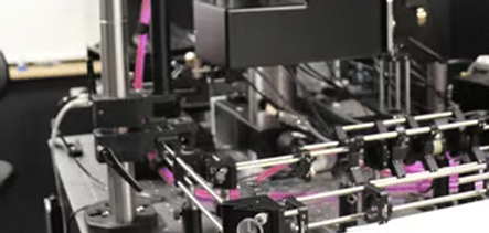
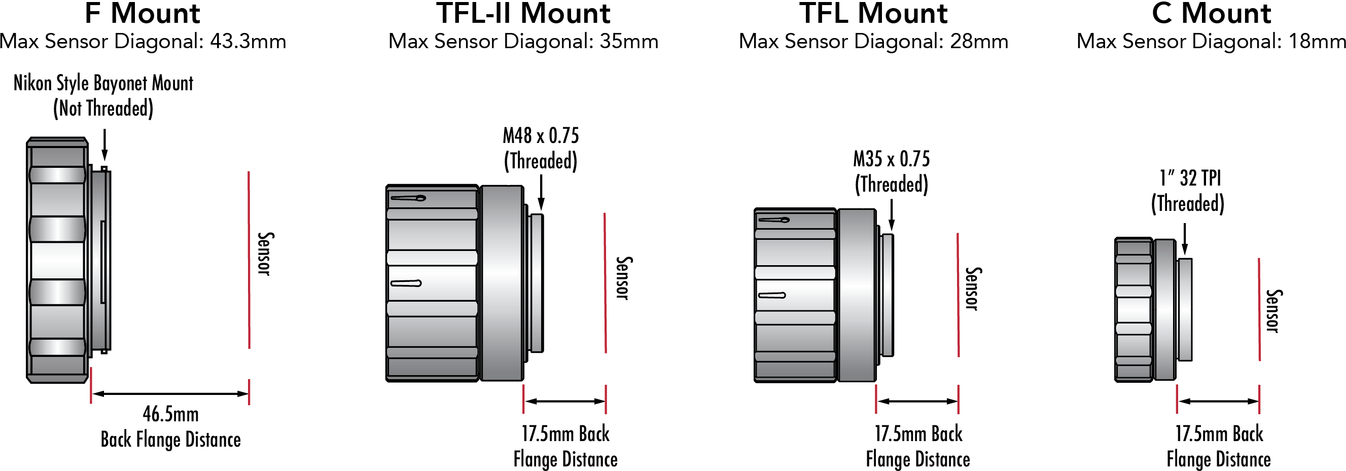
Assembling Compact Machine Vision Microscopy Systems with 120i Plan APO Infinity Corrected Objectives

Reduce the size and weight of your high magnification machine vision system with infinity corrected TECHSPEC® 120i Plan APO Infinity Corrected Objectives.

View Now Add to saved content

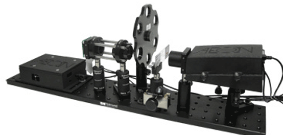
Choosing the Right Lasers and Optics for Ultrafast Microscopy

Join us for a discussion on selecting the best ultrafast laser sources and optical components for your ultrafast microscopy system to maximize performance.

View Now Add to saved content

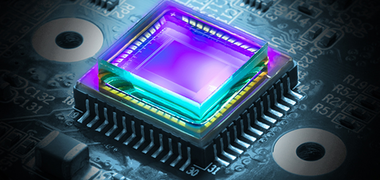
Ushering in a New Era of Edge AI with Sony AITRIOS and IMX500

WEBINAR: Join us for a 40-minute webinar presented by Sony Semiconductor Solutions Americas’ VP of Technology and Business Innovation, Mark Hanson, and Edmund Optics' Director of Imaging, Nicholas Sischka, to understand the nuance between AI, Edge AI, and Vision AI; what makes Sony AITRIOS unique; and how Lucid Vision Labs is putting the IMX500 sensor to action.

View Now Add to saved content

MICROSCOPY: Off-the-shelf components enable a new generation of confocal microscopy

View Now Add to saved content

Optical Microscopy Application: Fluorescence

Wondering how fluorescence microscopy works? Find out about the technique, systems, and more at Edmund Optics.

View Now Add to saved content

What's New In Optical Coatings: Applications in Life Sciences

Join Stephan Briggs, Biomedical Engineer at Edmund Optics, as he discusses tips for verifying filter performance.

View Now Add to saved content

Optical Microscopy Application: Phase Contrast

Think you know all the advantages for using phase contrast in optical microscopy? Advantages, image appearance, and technical details can be found at Edmund Optics.

View Now Add to saved content

Optical Microscopy Application: Brightfield Illumination

Looking to use a simple technique for optical microscopy? Check out this article on brightfield illumination to find out more at Edmund Optics.

View Now Add to saved content

Optical Microscopy Application: Darkfield Illumination

Darkfield illumination is the opposite of brightfield illumination. Find out how darkfield differs from brightfield in optical microscopy at Edmund Optics.

View Now Add to saved content

Optical Microscopy Application: Differential Interference Contrast

Differential interference contrast (DIC) is one of the polarization techniques that can be used in optical microscopy. Learn about this technique at Edmund Optics.

View Now Add to saved content

Fluorescence Microscopy: In-Line Illumination with Imaging Filters

Want to know about fluorescence microscopy? Read this article by a Biomedical Product Line Engineer at Edmund Optics to learn more.

View Now Add to saved content

Microscopy and the Analysis of a Trichrome Stain

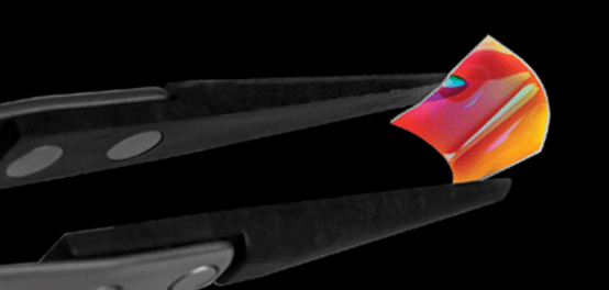
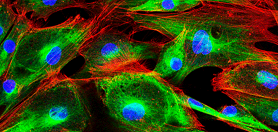
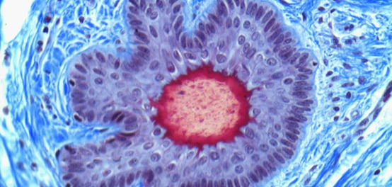
Imaging biological materials can be extremely difficult. Masson's trichrome stain helps differentiate components of cells from their surrounding tissues.

View Now Add to saved content

Portable Microscopy/Optics Innovations: High-performance, low-profile microscopy

View Now Add to saved content

Top 4 Trends of 2018

Advances in microscopy, imaging, and extreme ultraviolet optics

View Now Add to saved content

Vision Helps Reshape Microscopy, Biomedical Imaging Convergence

View Now Add to saved content

The right stuff: Lasers and optics for ultrafast microscopy

View Now Add to saved content

Non-Invasive In Vivo Imaging Using Fluorescent Microscopy

Edmund Optics and UVP developed a non-invasive In Vivo imaging technique using fluorescent microscopy.

View Now Add to saved content

Does Edmund Optics sell cameras with APS-C or APS-H sensors?

View Now Add to saved content

What's New In Optical Coatings: Introduction

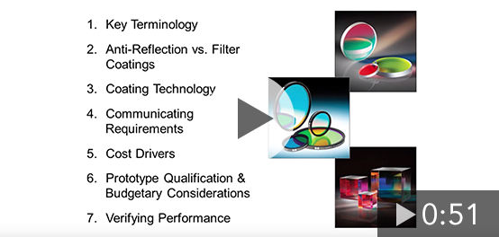
Join Stephan Briggs, Biomedical Engineer at Edmund Optics, as he provides an overview to his webinar including: Key Terminology, Coating Technology, Cost Drivers, and Application Examples.

View Now Add to saved content

What's New In Optical Coatings: Prototyping

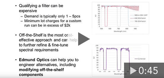
Join Stephan Briggs, Biomedical Engineer at Edmund Optics, as he discusses prototype qualifications including custom, stock and modified stock filter options.

View Now Add to saved content

Which imaging lenses feature a TFL Mount?

View Now Add to saved content